
Presentation
Flank pain
Patient Data


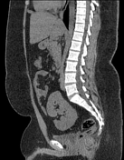
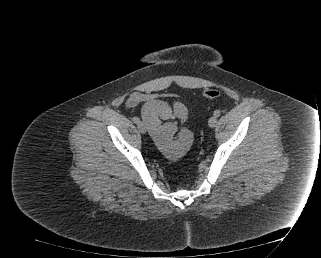
Both kidneys are not present at their normal anatomical location in renal fossa. Pelvic location of fused kidneys is noted with irregular shape and having bilateral renal vascular pedicles (both renal veins and arteries), in keeping with ectopic fused kidneys, known as pancake kidney.
There is a visualised renal pelvis facing left with a short ureter that crosses posteriorly draining at the right vesicoureteric junction. It is not possible to adequately assess ureters in the current non-contrast study with empty urinary bladder.
Right seminal vesicle is seen, but the left one is not identified in keeping with left seminal vesicle agenesis.
Prominent abdominal azygos vein, posterior to IVC.
Case Discussion
Both kidneys are ectopic and fused at the pelvic region (fused pelvic kidney), giving the characteristic appearance of a "pancake kidney". It is a rare renal fusion anomaly of the crossed fused variety. It is incidentally noted in this case and flank pain is likely not related to renal origin. Pancake kidney is usually located anterior to the aortic bifurcation. Seminal vesicle agenesis is an association with renal anomalies.
On current examination, there is only visualised renal pelvis and ureter, which is a more rare variant that has been previously reported in only four patients 1.
Differential diagnosis is solitary ectopic pelvic kidney, but it would have a single vascular pedicle.


 Unable to process the form. Check for errors and try again.