
Presentation
Recurrent pancreatitis now presenting during another acute episode
Patient Data
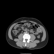

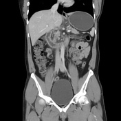

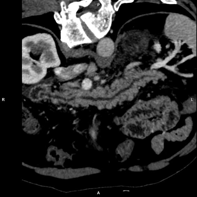
Mild generalised decrease in volume of pancreas for age. Pancreas is diffusely surrounded by fat stranding and oedema with area of mild non localised fluid in the lesser sac, right anterior pararenal fascia. Mild thickening of bilateral anterior pararenal fascia with few peripancreatic, portocaval and porta hepatis discrete enhancing subcentimetric lymph nodes. No areas of parenchymal pancreatic necrosis. No splenic artery aneurysm or splenic vein thrombosis. No pancreatic parenchymal calcification.
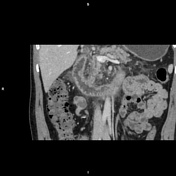
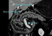
Independent dominant calibre of dorsal duct of Santorini draining separately into minor papilla. The ventral duct also joins the dorsal duct, whilst the CBD drains independently at the major papilla.

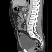
Curve planar reconstruction of the CT abdomen
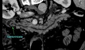
- Curved planar reconstruction of the dominant duct of Santorini entering independently in minor papilla with focal dilatation proximal to its duodenal insertion - santorinicele.
- Curved planar reconstruction of independent normal calibre common bile duct as it enters the major papilla.
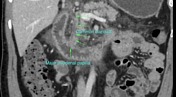

Curved planar reconstructions alond the pancreatic and common bile ducts with annotated anatomy
Case Discussion
Young patients with recurrent pancreatitis warrants work up to understand if there are any anatomical variations / developmental anomalies of the pancreatic ductal system especially in the absence of any form of biliary calcular disease. Interestingly this patient had a CT scan done 6 years ago and was reported as a case of mild acute pancreatitis. On careful review of the ductal system in the present scan, it was identified this patient has two independent drainage path for both pancreas and the rest of the biliary tree.
In events of idiopathic recurrent pancreatitis, pancreas divisum can be considered as an underlying aetiology. An MRCP may be done for confirmation and the gastroenterologist may opt for a therapeutic ERCP.


 Unable to process the form. Check for errors and try again.