
Presentation
Right flank pain.
Patient Data
Underwent CT stone protocol which ruled out urinary lithiasis.
There was, however, "fat stranding and fullness around the celiac trunk and SMA."
Contrast-enhanced CT was performed. Of note, the patient was given positive contrast per os, despite the radiologist's explicit instruction that water alone be given per os at the CT unit.
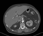

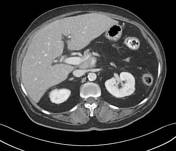

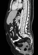
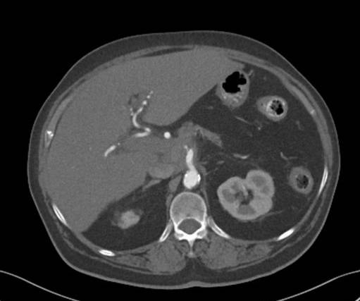
Pancreatic neck soft tissue mass slightly hypodense to the pancreas, completely encasing the celiac trunk and SMA as well as surrounding and narrowing the splenic vein and SMV near the portal confluence. The common bile duct and main pancreatic duct are not dilated.
Several small cystic dilatations along the main pancreatic duct, possibly IPMN. The pancreatic body and tail are atrophied.
Hepatic hilar lymphadenopathy measuring up to 22 mm (short axis) and hepatogastric ligament lymphadenopathy. No evidence of hepatic spread.
Findings are consistent with unresectable pancreatic adenocarcinoma.The celiac trunk is narrow (stenotic?) at its origin.
Bilateral elastofibroma dorsi.
Small cyst in each kidney.
The abdominal aorta shows soft and calcified plaque.


 Unable to process the form. Check for errors and try again.