
Presentation
Abdominal pain and postoperative swelling.
Patient Data
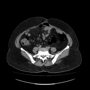

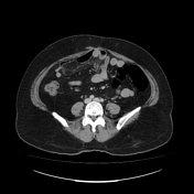
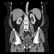
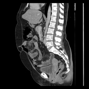

A complex lobulated cystic lesion with minimal internal septations and peripheral calcification is seen involving the pancreatic tail, measuring 1.9 x 1.7 cm. The rest of the pancreas appears normal.
Diastasis of the rectus abdominis is noted, with bulging of some abdominal organs anteriorly, such as the liver.
Small hernia defects are seen in the paraumbilical region with post-surgical changes.
Case Discussion
It is a type of mucinous cystic neoplasm of the pancreas and largely (~80%) occurs in the body or tail of the pancreas, and less frequently in the head of the pancreas (~20%).
The current case's CT scan shows classic features of a mucinous tumor, including a round/oval contour, peripheral calcification, and a few internal septations.


 Unable to process the form. Check for errors and try again.