
Presentation
Abdominal pain, known diverticulosis - diverticulitis?
Patient Data

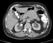
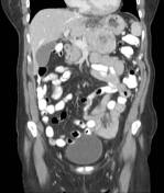

Diverticula along the colon, numerous in the descending colon and innumerable in the sigmoid colon. No sign of acute inflammation.
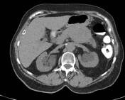
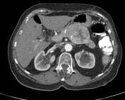
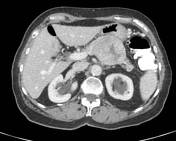
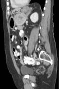
Ovoid mass in the body of the pancreas measuring 7.4 x 5.7 x 5.4 (RL x AP x CC) cm with central stellate hypodensity, the very center of which represents necrosis. The pancreatic artery passes through the mass and is not compressed or distorted by it. The mass compresses the pancreatic vein. The pancreatic tail is atrophic and the main pancreatic duct through it is dilated. Prominent vessels in the hepatogastric ligament.
Several small cystic lesions in the left hepatic lobe, the largest in segment 4b, measuring 2.4 cm in length. Irregular 10-mm focus of arterial enhancement in hepatic segment 7 - vascular shunt?
Numerous bilateral peripelvic cysts.
Case Discussion
Tumor markers were normal.
Underwent distal pancreatectomy and splenectomy.
Histopathology report:
Pancreatic well-differentiated neuroendocrine tumor, G2, confined to the pancreas
Tumor size 6.2 cm. Mitotic rate - 3 per 10 HPF (2 mm2). Ki67 - 10%. Proximal pancreatic, anterior and posterior margins are free of tumor. Angiovascular invasion not identified. Fourteen (14) peripancreatic lymph nodes are free of tumor. Unremarkable spleen.
Comment: Immunohistochemical stains for chromogranin and synaptophysin are positive.
pTNM: pT3, N0.


 Unable to process the form. Check for errors and try again.