
Presentation
A 77 year old gentleman presented to our outpatients department with a 7 year history of a painless enlarging right sided neck lump. He was otherwise asymptomatic. His significant past medical history included asthma, and he was an ex-smoker. MRI indicated the tumor was indeed a benign lipoma. He is currently being followed up in outpatients.
Patient Data
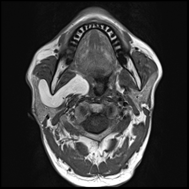
A 5.7 x 6.5 x 3.5cm lipomatous lesion can be seen deep to the parotid and in the parapharyngeal soft tissues on the right side.
Case Discussion
Primary tumors of the parapharyngeal space are extremely are and compromise 0.5% of all head and neck masses (1). Typically, these lipomas present as painless intraoral or neck lumps. However, depending on their size and location they can present with neurovascular compromise (2). An important differential to rule out is liposarcoma (3). Management of parapharyngeal space lipomas include, appropriate radiological investigations, including MRI, biopsy of the lesion if possible, and surgical excision of the lipoma (1,3). Conservative management with close long-term follow is possible if liposarcoma has been ruled out (1,3). Our patient was managed with the latter option.


 Unable to process the form. Check for errors and try again.