
Presentation
Abdominal pain. Ultrasound reveals a lesion in the left liver suspected to be a bile duct tumour.
Patient Data
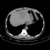

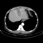
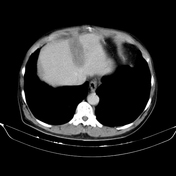
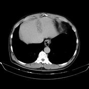

Lesions in segment II and IVA of the liver show decreased density on non-contrast imaging, measuring 32 x 66 x 41 mm, with ill-defined borders. There is poor enhancement in both the arterial and venous phases, with surrounding liver parenchyma exhibiting perfusion abnormalities. Delayed enhancement is observed (enhancement of the internal septations), and the cluster sign is also clearly visible.
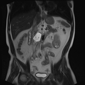
The right kidney contains a calcified cyst (Bosniak class IIF), while the left kidney has several simple cysts (Bosniak class I).
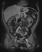
A diverticulum is seen in the D3 segment of the duodenum.
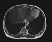
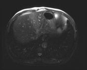
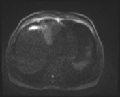
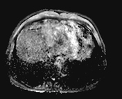
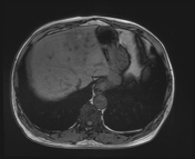
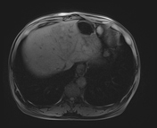
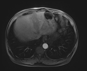
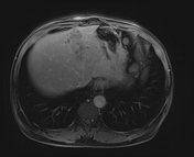
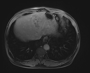
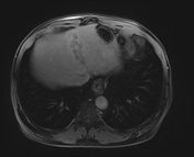

A liver lesion in segment II, measuring 32 x 66 x 41 mm, and several small lesions in the adjacent segment IVA, with heterogeneous high and low signal intensity on T1FS, high signal on T2W and STIR, partially restricted diffusion, surrounding perfusion abnormalities, poor enhancement after contrast injection, with delayed enhancement (mainly in the internal septations), cluster sign (+), and no mass effect observed.
A few renal cysts in both kidneys, with some cysts showing high signal intensity on T1FS (Bosniak Class IIF).
Diverticulum in the D3 segment of the duodenum.
Blood test results:
eosinophil count: 3.4 K/µL (elevated)
eosinophil percentage: 37.6% (elevated)
CA 19-9 quantification (Carbohydrate Antigen 19-9): 10.3 (normal)
Echinococcus granulosus autoimmune antibody: positive
Fasciola autoimmune antibody: positive
Schistosoma autoimmune antibody: positive
Trichinella spiralis autoimmune antibody: positive
Clonorchis/opisthorchis autoimmune antibody: positive
HCV Ab rapid test: negative
HBsAg autoimmune test: negative
Case Discussion
The initial imaging findings suggest a hepatic abscess. A differential diagnosis includes intrahepatic cholangiocarcinoma (bile duct tumour).
After correlating with the laboratory results, the final diagnosis is parasitic liver abscess.
The patient was treated with antiparasitic medication, leading to improvement in clinical symptoms and blood test results.


 Unable to process the form. Check for errors and try again.