
Presentation
Four-year history of asymptomatic hypercalcaemia and hypercalciuria associated with hyperparathyroidism being managed medically. Seen by endocrinology for follow up of her hyperparathyroidism.
Patient Data
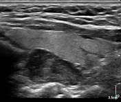
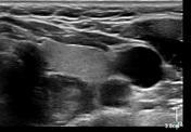
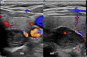
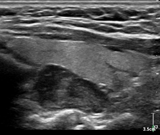
Hypoechoic mass measuring 2.5 x 1.5 x 1.1 cm immediately deep to the left thyroid lobe. Colour doppler images demonstrate internal vascularity.
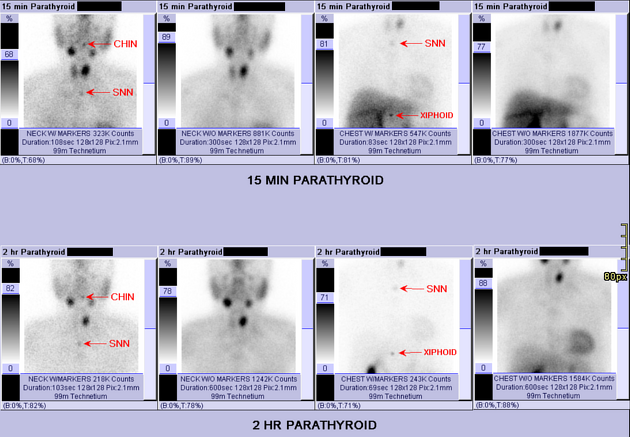
Avid radiotracer uptake over the region of the left thyroid on immediate images which persists on 2 hour delayed images.
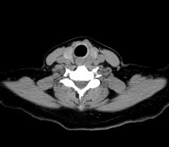
Isodense nodule measuring 1.3 x 0.9 cm posterior to the left thyroid.

Physiologic mild bilateral thyroid radiotracer uptake with intense focal radiotracer uptake in the soft tissue nodule posterior to the left thyroid lobe.
Case Discussion
A classic example of a parathyroid adenoma in a patient with a history of asymptomatic hyperparathyroidism. Initially visualised with ultrasound, and subsequently seen on scintigraphy and SPECT/CT.


 Unable to process the form. Check for errors and try again.