
Presentation
Left preauricular neck swelling.
Patient Data
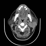

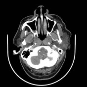

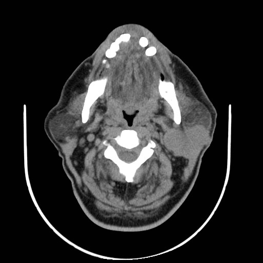
An irregular infiltrating soft tissue density lesion originating from left parotid gland and extending medially displacing the left carotid sheath. It has a heterogeneous enhancement in the post I.V contrast phase.
Different sizes cervical lymph nodes are noted in the neck, the largest is located in left anterior triangle superiorly and also in the left supra-clavicular angle.
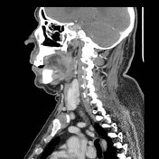
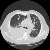
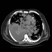
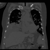
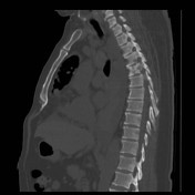
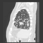
Well defined variable sizes and shape focal metastatic lesions are seen within both lung fields. Evidence of right sided mild to moderate pleural effusion with possible underlying consolidation /collapse.Evidence of mediastinal lymphadeopathy are seen. Some show calcific foci. Bony osteolysis and deposits of D3 and D5 vertebrae.
Case Discussion
The case is a histopathologically proven high grade mucoepidermoid parotid carcinoma.
It can cause pulmonary and vertebral osseous deposits.


 Unable to process the form. Check for errors and try again.