
Presentation
Left orbital swelling, pain, tearing, redness, and vision loss.
Patient Data
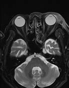

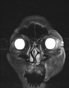
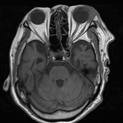
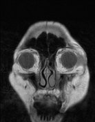
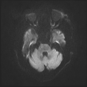
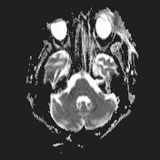
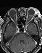
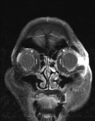

Swelling, oedema, and thickening of the soft tissues anterior to the left orbital septum, extending to the lateral cheek region, with increased T2FS/FLAIR signal, diffusion restriction, and heterogeneous contrast enhancement. A small fluid collection is present in the preseptal orbital space.
The bilateral globes, extraocular muscles, and optic nerve sheaths are normal. No inflammatory changes in the intraorbital fat bilaterally.
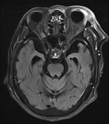
Right corona radiata lacunar infarct.
Suspected left temporomandibular joint arthritis is also noted.
Case Discussion
Blood tests revealed elevated white blood cell count, neutrophils, and CRP levels. The imaging findings, clinical symptoms, and laboratory results are consistent with periorbital cellulitis.
Orbital infections can be classified according to Chandler's classification of orbital infections. This case represents type I, as the infectious changes are confined to the preseptal space.
The patient was treated with oral antibiotics, resulting in significant improvement in clinical symptoms and blood test findings.


 Unable to process the form. Check for errors and try again.