
Presentation
Presented with mass at the left forearm (volar surface)
Patient Data
Age: 50 years
Gender: Female
From the case:
Peripheral nerve sheath tumour


Show annotations
Download
Info
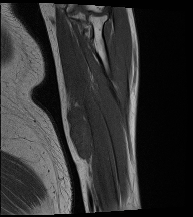
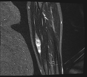
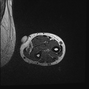
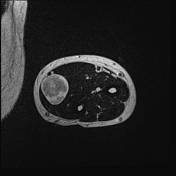
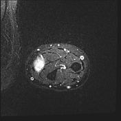
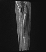
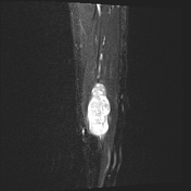
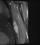
There is a well-defined, oval-shaped mass measuring about 56 x 18 mm in the course of the median nerve branch of the left forearm.
The mass shows homogenous T1 hypointensity and heterogenous T2 hyperintensity with a discreet but visible split fat sign.
Case Discussion
Appearance is suggestive of a peripheral nerve sheath tumour of the median nerve.


 Unable to process the form. Check for errors and try again.