
Presentation
Intellectual disability and seizures.
Patient Data
Age: 8 years
Gender: Male
From the case:
Periventricular leukomalacia


Download
Info

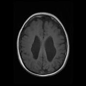
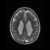
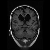
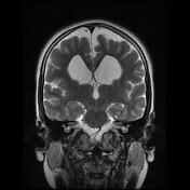
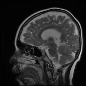
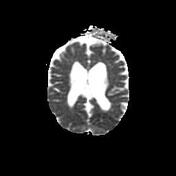
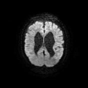
The MRI sequences demonstrate:
ventriculomegaly with wavy and irregular lateral contours
loss of periventricular white matter with increased signal on FLAIR and T2 sequences
thinning of the corpus callosum
Case Discussion
MRI features are most consistent with end-stage periventricular leukomalacia (PVL).


 Unable to process the form. Check for errors and try again.