
Presentation
Handicapped patient with cerebral palsy presented for follow up MRI.
Patient Data
Age: 16 years
Gender: Male
From the case:
Periventricular leukomalacia


Show annotations
Download
Info

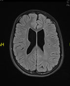
Mildly dilated lateral ventricles with irregular outline and subtle periventricular gliotic changes.
Diminished periventricular white matter.
Mega cisterna magna.
Case Discussion
Periventricular leukomalacia (PVL) is a form of brain injury that affects infants, particularly premature babies. It involves the necrosis of the periventricular white matter. This condition is most commonly associated with premature birth and is a leading cause of cerebral palsy and other neurological impairments.
MRI features consistent with end-stage periventricular leukomalacia (PVL).


 Unable to process the form. Check for errors and try again.