
Presentation
Chronic progressive left ear hearing loss, No other clinical findings.
Patient Data
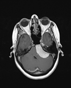

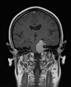
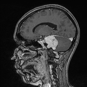
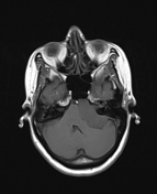
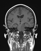
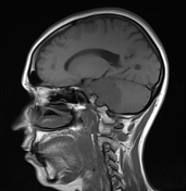
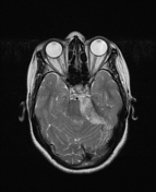
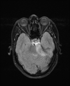
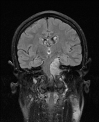
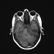
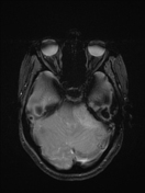
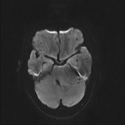
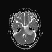

Well-defined extra-axial infratentorial space-occupying lesion, measuring about 4*3.8*2.5 cm, attached to dura in the left petroclival region with obtuse angles at dural margins, extending into the left internal acoustic canal, isointense on T1 and hyperintense on T2 and FLAIR, with avid homogeneous enhancement on post-contrast T1 sequences, and high ADC value indicating high diffusion.
A mass effect is noted on the medial temporal lobe, brainstem, left cerebellar hemisphere and the 4th ventricle, without causing hydrocephalus. Minimal surrounding edema is noted.
Signs of extra-axial mass are noted:
dural-based lesion (dural tail sign)
widing of adjucent CSF spaces (meniscus sign) and (CSF cleft sign)
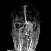
vessels pushed inward (displaced vessels)
inward bowing of the gray-white matter junction (white matter bulking sign)
The previous findings indicate Petroclival meningioma
Pathological report:
Sections show neoplastic meningothelial cell proliteration within prominent fibrocollagenous background. Tumor cells are arranged in syncytial and whorling patterns. Occasional psammoma bodies are noted. There is no evidence of significant nuclear atypia or increased mitotic activity.
Case Discussion
Imaging findings correlate with petroclival meningioma.
The diagnosis was confirmed by pathology (Meningioma grade 1).


 Unable to process the form. Check for errors and try again.