
Presentation
Long-lasting headaches. Arm and leg weakness.
Patient Data


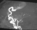


In the posterior part of the right frontal lobe is a cluster of abnormal vessels with marked aneurysmal dilatation. The feeding artery is the right middle cerebral artery. It connects directly to the largest aneurysmal dilatation. This directly drains into the superior sagittal sinus and inferior sagittal sinus without a visible intervening nidus. A region of blood product is present inferiorly consistent with prior haemorrhage.
Case Discussion
Pial arteriovenous fistulae (pAVF) are rare vascular malformations that usually consist of a single dilated pial artery connecting directly to an enlarged cortical draining vein. In this case, we found a dilated right middle cerebral artery that connects directly to the superior sagittal sinus. We do not see a "nidus," so we do not think it is an arteriovenous malformation (AVM).
Many types of cerebrovascular lesions occur in the central nervous system, each receiving a different name according to its morphological features. If there are abnormal vessels between the arterial and venous systems, they are called a nidus and are considered an AVM. If there is no nidus, the condition is referred to as an arteriovenous fistula (AVF), which can be pial or dural. If the blood vessels are not related to the dura mater and come from the cortical and/or pial regions, they are called 'pial arteriovenous fistulae' or pAVF.
Even though the vast majority of patients with pAVF are treated with an endovascular approach, the choice of treatment should be multidisciplinary and based on the experience of the medical team. It is important to evaluate the location and degree of lesion complexity.
A cerebral angiogram confirmed an arteriovenous fistula with the right middle and anterior cerebral arteries as feeding vessels and the superior cerebral veins and superior and inferior sagittal sinus as draining vessels, which was subsequently treated by endovascular coiling.


 Unable to process the form. Check for errors and try again.