
Presentation
Headache and double vision (diplopia).
Patient Data


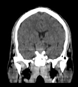
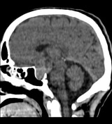
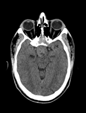
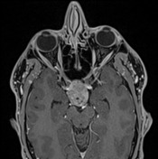
A large mass is seen within sellar fossa and suprasellar cistern and shows cystic changes without obvious calcification or haemorrhagic component.
No obvious bony destruction.

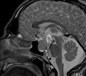
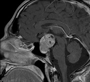
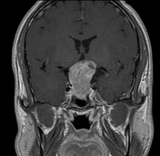

There is a large sellar mass extending to the suprasellar area (giving a snowman appearance) and causing the expansion of the sella and mass effect to the floor of 3rd ventricle superiorly.
The lesion appears isointense on T1W sequence and slightly hyperintense on T2W sequences, enhancing heterogeneously on post-contrast exam and showing cystic changes, without obvious calcification or haemorrhagic component.
The optic chiasm is not clearly visualised being mostly compressed by the lesion
Case Discussion
Pituitary macroadenomas are adenomas over 10 mm in size. They appear as soft tissue lesions with areas of necrosis as they get bigger along with upward growing (suprasellar extension and compression of the optic chiasm).
The snowman configuration is noted in this case and is a useful sign to help distinguish between a pituitary macroadenoma and a meningioma.


 Unable to process the form. Check for errors and try again.