
Presentation
Shortness of breath with a medical history of congestive heart failure, diabetes, and hypertension.
Patient Data
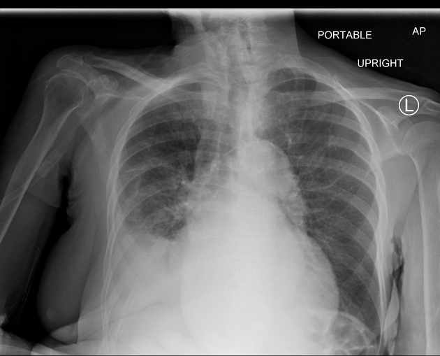
Moderate atherosclerotic disease at the aortic arch.
Large cardiac silhouette. Right lower lobe interstitial opacity. Blunted right costophrenic angle may represent pleural effusion. Left lung shows linear opacities at the bases may represent atelectasis.
Right lower lobe infiltration with right pleural effusion suspicious for pneumonia.
Case Discussion
This represents a case of pleural effusion (PF), defined as the accumulation of fluid between the parietal and visceral pleura.1
PF may be due to several underlying diseases, including heart failure, cancer, pulmonary embolism, and infection.2
In this case, laboratory findings revealed an elevated white count (13.5 x10³/mcL).
A combination of cefepime and azithromycin used for treatment improved her condition.


 Unable to process the form. Check for errors and try again.