
Presentation
Monoparesis of the left lower limb.
Patient Data


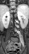


Moderate lumbar scoliosis of left-sided convexity with enhancing plexiform masses extending along the distribution of the left L2, L3, and L4 nerve roots, enlarging the neural exit foramina from L2-L3 to L4-L5 levels. Moderate vertebral scalloping is noted. Atrophied ipsilateral psoas and gluteal muscles.
The brain MRI was normal (not shown).
Case Discussion
MRI features of plexiform neurofibromas along the left L2, L3 and L4 nerve roots in a patient not known for neurofibromatosis type 1 (probably isolated plexiform neurofibromas).
Plexiform neurofibromas are benign tumours of peripheral nerves (WHO grade I), arising from a proliferation of all neural elements. They are seen in approximately 30% of patients with NF1.


 Unable to process the form. Check for errors and try again.