
Presentation
Progressive painless swelling of the left submandibular space
Patient Data


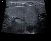
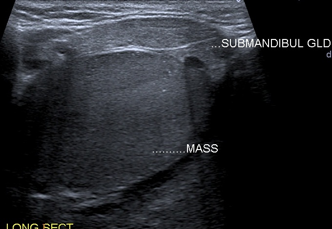
A well-defined left submandibular cystic mass with thin wall and homogeneous echogenic content, posteromedial to the ipsilateral submandibular gland which appears of normal size and echotexture but displaced anterolaterally. The right submandibular gland is of normal size and echotexture.


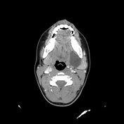
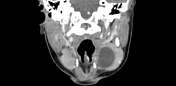
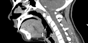
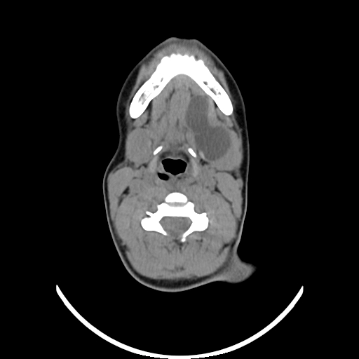
The mass is well-circumscribed, of cystic nature with no peripheral enhancement, located in the left sublingual space between the mylohyoid muscle laterally, geniohyoid/genioglossus complex medially, and the ipsilateral submandibular gland posterolaterally with extension to the adjacent submandibular space.
Case Discussion
Ultrasound and CT features are typical of a plunging ranula.


 Unable to process the form. Check for errors and try again.