
Presentation
Shortness of breath.
Patient Data

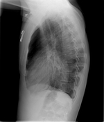
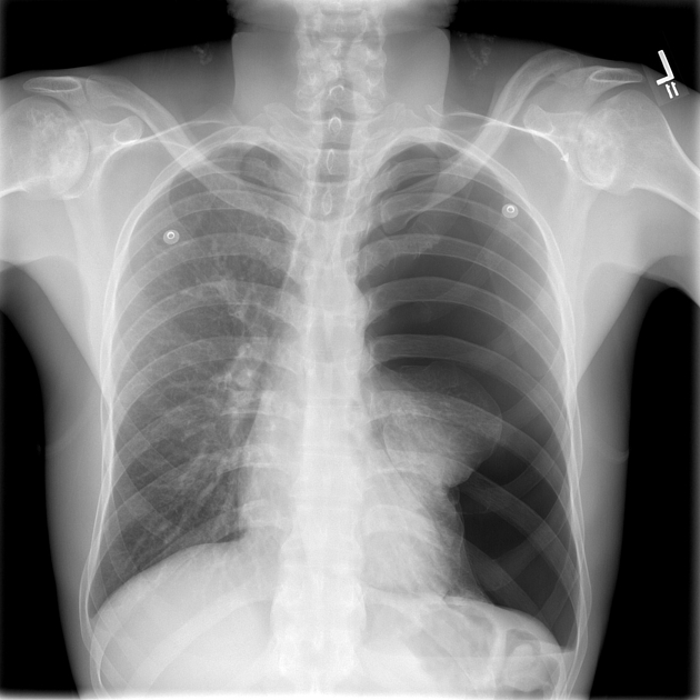
Large left pneumothorax with passive atelectasis left upper and left lower lobes. Mediastinal shift to the right. Lungs grossly clear. Patchy lucency and sclerosis both humeral heads likely avascular necrosis.
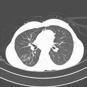

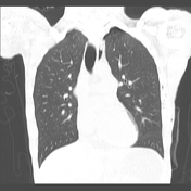
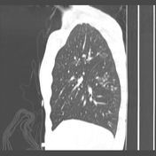

Left pleural catheter enters the anterior left lung base along an accessory fissure, with the catheter parallelling the left lateral aspect of the mediastinum adjacent to the aorta and pulmonary artery, tip abutting the left innominate vein. Trace left pneumothorax, seen primarily at the left lung apex. Small left apical bulla.
Moderate amount of fluid and debris within the oesophagus. Small airways nodules within the superior segment of the right lower lobe, and posterior segment of the right upper lobe, in keeping with infectious or aspiration related bronchiolitis. No pleural effusion or consolidation.
Case Discussion
One of the causes of a spontaneous pneumothorax is rupture of an apical bulla: that was the likely mechanism in this case.


 Unable to process the form. Check for errors and try again.