
Presentation
Incidental finding.
Patient Data
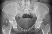
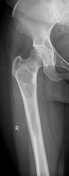
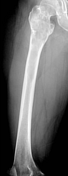
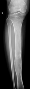
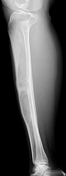

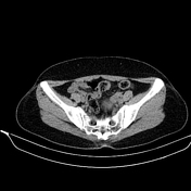
On the x-ray images of the pelvis, femur, and right tibia, multiple bone lesions were noted in the superior pubic ramus, the upper third of the femur, the acetabulum, and the upper two-thirds of the tibia. These lesions are lytic and centred in the medulla, causing thinning and mild expansion of the cortex. The matrix has a ground-glass appearance with no periosteal reaction. The transition zone is narrow with a thin sclerotic rim surrounding the lesions and there are no pathological fractures.


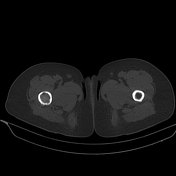
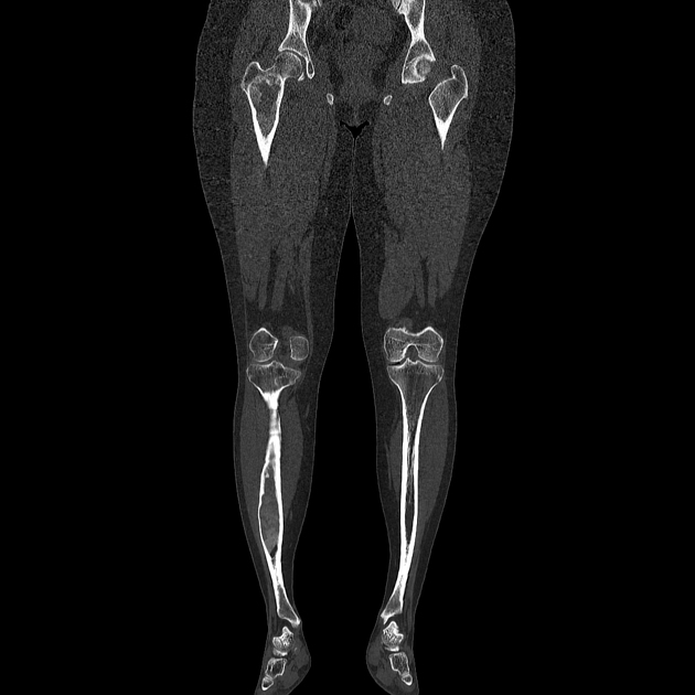
CT helps to clearly visualise the lesions described on x-ray, including:
well-defined borders
expansion of the bone
endosteal scalloping
no periosteal reaction
no pathological fractures
no soft tissue mass surrounding

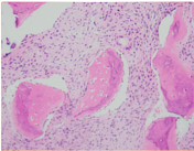
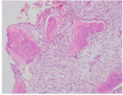
Biopsy location: proximal tibia.
Microscopic description: the histological description shows a combination of sparsely cellular fibrous tissue with small trabecular bone fragments interspersed, lacking a discernible lining of osteoblasts.
Conclusion: fibrous dysplasia.
Case Discussion
The patient underwent whole-body bone scintigraphy with Tc99m-MDP, which revealed abnormal increased radiotracer uptake in the superior ramus of the right pubic bone, the upper third of the right femur, and the upper two-thirds of the right tibia. Radiotracer uptake in the soft tissues was normal and physiologically appropriate. The kidneys were clearly visible on the scan. Fibrous dysplasia is metabolically active and should not be mistaken for malignancy.
The imaging and histopathological findings confirm the diagnosis of polyostotic fibrous dysplasia.
This is an incidental case, with no pathological fractures, no clinical symptoms, no significant bone deformities, and no signs of malignant transformation. Management is focused on maintaining bone quality through dietary measures and exercise, with the prevention of secondary fractures being emphasised.


 Unable to process the form. Check for errors and try again.