
Presentation
Fall. Hyperflexion injury.
Patient Data


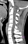

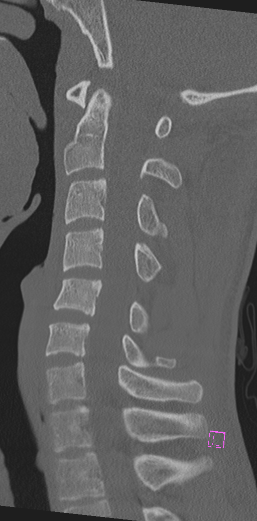
No fractures. Focal widening of the C4/C5 interspinous space is concerning for ligamentous injury and further assessment with MRI is recommended.
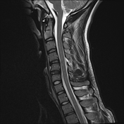
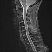
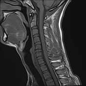
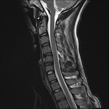
Focal widening of the interspinous space posteriorly at C4/C5 is associated with a prominent focal high T2 signal and disruption of the ligamentum flavum and probably also of the interspinous ligament. The supraspinous ligament/nuchal ligament appears intact. The disc has high T2 signal within the annulus fibres posterolaterally on the left with only a small protrusion and no canal stenosis.
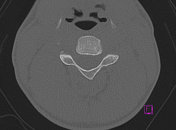
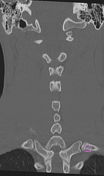
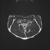
No cord compression or abnormality. The posterior longitudinal ligament and anterior longitudinal ligaments are intact. The remainder of the cervical spine is unremarkable.
Conclusion:
C4/C5 posterior tension band injury (disruption of ligamentum flavum and interspinous ligaments) with injury to the disc annular fibres, posterolaterally on the left; AO = B2.


 Unable to process the form. Check for errors and try again.