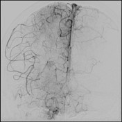
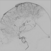
Proximal right MCA M1 segment embolic occlusion
Diagnosis certain
Updates to Study Attributes
Findings
was changed:
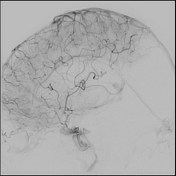
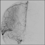
Baseline frontal DSA projection of the right internal carotid artery demonstrating complete occlusion of the proximal right MCA M1 segment and retrograde filling of distal MCA branches from ACA-MCA pial communications. Baseline lateral DSA projection demonstrates retrograde filling of distal MCA branches from ACA-MCA pial communications.
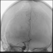
Frontal and lateral right ICA DSA withshow stent-retriever in situ (Stryker TREVO XP 6 x 25 mm) highlighting the clot and providing "temporary endovascular bypass".
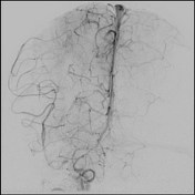
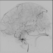
Final DSA post retrievalfollowing removal of the stent-retriever demonstrating re-perfusionreperfusion of the previously occluded MCA. TICI 2C angiographic result (some underfilling of small distal parietal MCA cortical branches).
modified TICI 2C
Images Changes:
Image DSA (angiography) (Internal carotid artery) ( update )
Description
was removed:
Image DSA (angiography) (Internal carotid artery) ( update )
Description
was removed:
Image DSA (angiography) (Internal carotid artery) ( update )
Description
was removed:
Image DSA (angiography) (Internal carotid artery) ( update )
Description
was removed:
Image DSA (angiography) (Internal carotid artery) ( update )
Description
was removed:
Image DSA (angiography) (Internal carotid artery) ( update )
Description
was removed:
Image DSA (angiography) (Internal carotid artery) ( update )
Description
was removed: