
Presentation
History of surgery for femoral fracture 4 weeks prior, presented with dyspnoea, cough and left thoracic pain.
Patient Data


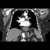
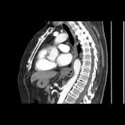

Filling defects of the left main pulmonary artery extending into the lobar arteries and their segmental branches mainly in the left lower lobe with thin rim of contrast around the central filling defect "polo mint sign"
Filling defects of the dorsal branch of the right upper lobar artery and right lower lobar artery and its segmental branches.
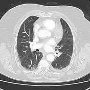
Minimal left pleural effusion with triangular shaped peripheral pulmonary opacities of the lower lobe (result of lung infarction secondary to pulmonary embolism). Dilated left atrium with mitral valve calcification.
Sliding hiatal hernia is noted.
Case Discussion
CT features of an acute pulmonary embolism complicated by lung infarct (the D-dimer level was at 4530 ng/mL in this case).


 Unable to process the form. Check for errors and try again.