
Presentation
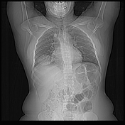
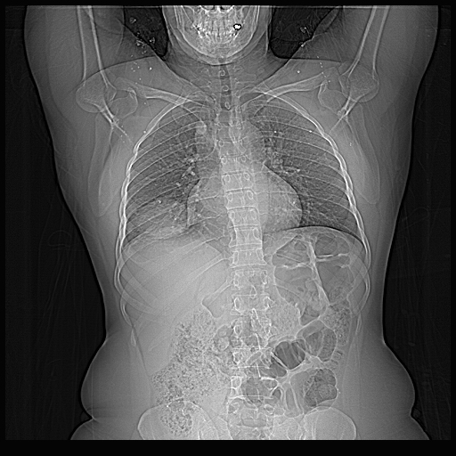
Chronic cough. A chest radiograph shows a rounded opacity in the right lower zone.
Patient Data
Age: 40 years
Gender: Female
From the case:
Pulmonary hydatid cyst

Show annotations
Download
Info
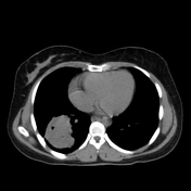
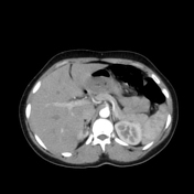
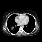
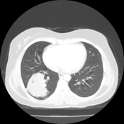
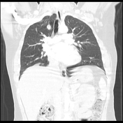
Mass in the right lower lobe with inner fluid density associated with scattered foci of air densities.
An incidental azygos lobe is denoted by the azygos vein crossing through the right upper lobe to join with SVC.
Case Discussion
The findings are suggestive of an infective pulmonary hydatid cyst, with the clinical presentation, including a history of exposure to Echinococcus, providing further support.
The presence of scattered foci of gas within the cyst raises the possibility of an infective or ruptured hydatid cyst.
An incidental azygos lobe and vein are noted.


 Unable to process the form. Check for errors and try again.