
Presentation
Road traffic accident
Patient Data
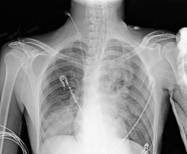
Bilateral pulmonary ground glass opacities, mainly in right lower zone and left upper zone.
Endotracheal tube and right central vein line are noted.
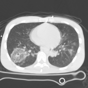


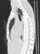
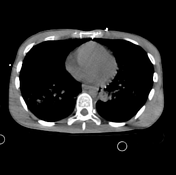
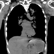

Multiple oval-shaped cavitary lesions with air/fluid level surrounded by air space opacities are seen in anterior and apical segments of left upper lung lobe and right lower lung lobe, features are suggestive of pulmonary lacerations.
High position of endotracheal tube.
Case Discussion
There are different patterns of lung parenchymal injury, e.g pulmonary contusion, pulmonary haemorrhage and pulmonary laceration. The latter usually appears as a round or oval-shaped cavity filled with blood and/or air. This is due to the pulmonary elastic recoil, that makes lung tissue pulls away from a laceration creating a cavity.
The patient developed bilateral pleural effusions next day that exacerbated the condition and aggravated respiratory failure and unfortunately he died.


 Unable to process the form. Check for errors and try again.