
Presentation
Rushed into the ED with sudden dyspnea and left sided chest pain.
Patient Data
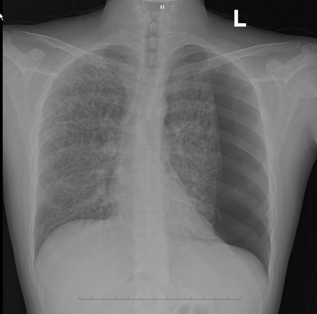
Bilateral coarse reticular interstitial pattern with a large left pneumothorax and mediastinal shift to the right.


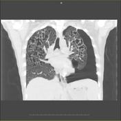

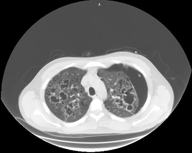
Computed tomography scan of the chest showed numerous thin-walled pulmonary cysts of varying sizes and shapes, some of which are confluent and result in large cystic lesions. There is more middle and upper lobe predilection.
Case Discussion
Diagnosis was confirmed by bronchoscopic biopsy.
Langerhans’ cell histiocytosis (LCH) is a group of disorders characterized by infiltration of multiple systems by Langerhans’ cell. The exact pathophysiology of this idiopathic disorder is unknown. Reticulo-micronodular infiltration is the commonest pattern observed by CXR, although the abnormalities vary with the stage of the disease 1.
CT, especially the HRCT plays an important role in the diagnosis of of PLCH 2 . The typical HRCT pattern of PLCH is thick- and thin-walled cysts combination with small poorly limited nodules, cavitated nodules, which affect both the peripheral and the central parts of the lung fields 1.


 Unable to process the form. Check for errors and try again.