
Presentation
Reduced oxygen saturations, bilateral crepitations and reduced air entry on auscultation bilaterally. Three week history of worsening shortness of breath on exertion and bilateral pedal oedema.
Patient Data

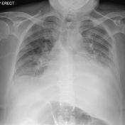

There is perihilar air space opacification seen bilaterally with a small left pleural effusion. Appearances are suggestive of pulmonary oedema.
Case Discussion
This patient presented with reduced oxygen saturations to the emergency department after a 3 week history of progressive dyspnoea and was diagnosed with acute pulmonary oedema secondary to heart failure, which was confirmed via echocardiogram. They were successfully treated with IV furosemide.
Some classic features of pulmonary oedema are demonstrated on this chest x-ray: pleural effusion and perihilar opacification. Other common features include upper lobe diversion and cardiomegaly (heart size was unable to be accurately assessed on these films).


 Unable to process the form. Check for errors and try again.