Pulmonary sequestration (prenatal and postnatal ultrasound)
Updates to Study Attributes
An echogenic lesion is noted in left lower hemithorax posteriorly. Its base is noted abutting spine. LesionThe lesion also abuttsabuts the lower thoracic thoracic aorta, part of oesophagus and heart.
Lesion The lesion is posterior and close to normal appearing-appearing stomach bubble. No defect is noted in adjacent spine.
Lesion The lesion appears to extend in the upper abdomen posterior to stomach. There seems to be a defect in the left hemidiaphragm. Lesion receives arterial supply from the aorta.
One or probably two arterial are seen supplying the lesion. It is measuring Cranio-caudal - 15 mm
Axial , Axial - 10 mm
and Antero-posterior - 10 mm.
No cystic spaces are noted in the lesion.
No hydrothroax No hydrothorax is noted.
No No mass effect is noted on heart or stomach bubble.
RIGHT hemidiaphgramThe right hemidiaphragm is normal.
RIght The right lung and rest of the left lung are normal.
Lesion receives arterial supply from aorta. One or probably two arterial are seen supplying the lesion.
Mild fullness of LEFTleft renal pelvis is noted.
No No other abnormality could be localized in fetus.
Image Ultrasound ( update )

Image Ultrasound (Transverse) ( update )
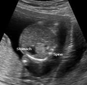
Image Ultrasound (Transverse) ( update )

Image Ultrasound (Longitudinal) ( update )

Image Ultrasound (Longitudinal) ( update )

Image Ultrasound (Coronal) ( update )

Image Ultrasound (Longitudinal) ( update )

Image Ultrasound (Transverse) ( update )

Image Ultrasound (Transverse) ( update )

Image 9 Ultrasound ( create )
Updates to Study Attributes
An echogenic lesion is noted posterior to gastro-oesophageal junction.It. It also extends to lower left hemithorax.
Lesion abutts Lesion abuts the upper abdominal aorta. LesionThe lesion is posterior to heart.
Discontinuity is noted in left hemidiaphragm. Left adrenal gland is normal.
Lesion The lesion is echogenic without cystic changes.
NO pleural effusion is noted in either of pleural cavities.
Lesion Lesion receives arterial supply from aorta. One artery is seen supplying the lesion. Discontinuity is noted in left hemidiaphragm. Left adrenal gland is normal.
No pleural effusion is noted in either of pleural cavities.
Hydronephrosis without parenchymal loss is noted in LEFTthe left kidney with a distended bladder. No hydroureter is noted..
No other abnormality could be localized in the abdomen.
Updates to Case Attributes
Other differentialdifferentials for an echogenic lung lesion for antenatal ultrasound includesinclude CCAM ( congenital(congenital cystic adenomatoid malformation), lobar emphysema, bronchial atresia; all supplyedsupplied by pulmonary artery.
Antenatal and postnatal Findings of lower chest echogenic mass with extension below diaphramdiaphragm along with blood supply from aorta favours the diagnosis of pulmonary sequestration.
-<p>Other differential for an echogenic lung lesion for antenatal ultrasound includes CCAM ( congenital cystic adenomatoid malformation ), lobar emphysema, bronchial atresia; all supplyed by pulmonary artery.</p><p>Antenatal and postnatal Findings of lower chest echogenic mass with extension below diaphram along with blood supply from aorta favours the diagnosis of <strong>pulmonary sequestration</strong>.</p>- +<p>Other differentials for an echogenic lung lesion for antenatal ultrasound include CCAM (congenital cystic adenomatoid malformation), lobar emphysema, bronchial atresia; all supplied by pulmonary artery.</p><p>Antenatal and postnatal Findings of lower chest echogenic mass with extension below diaphragm along with blood supply from aorta favours the diagnosis of <strong>pulmonary sequestration</strong>.</p>