
Presentation
Haemoptysis.
Patient Data
Age: 45 years
Gender: Male
From the case:
Pulmonary tuberculosis


Download
Info
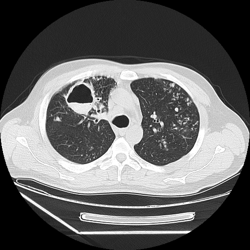
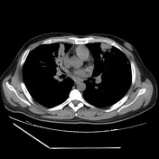
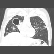
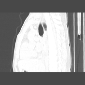
Multiple thick-walled cavitary lesions are seen within areas of consolidation, the largest cavitary lesion is in the anterior segment of the right upper lobe with an air-fluid level.
Scattered well-defined nodules and centrilobular nodules in a linear branching pattern "tree-in-bud appearance" indicating extensive endobronchial spread.
Diffuse ill-defined patchy consolidations.
Case Discussion
This case demonstrates features of pulmonary tuberculosis in a sputum smear-positive TB patient.


 Unable to process the form. Check for errors and try again.