
Presentation
Acute confusion and fever.
Patient Data
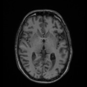

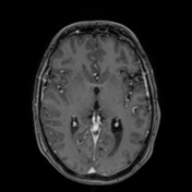
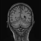
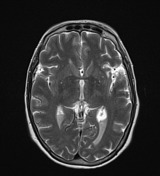
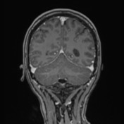
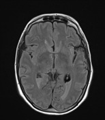
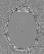
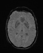

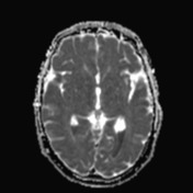
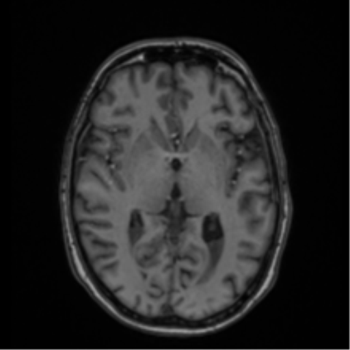
There is dependent material within the occipital horns of the lateral ventricles that is hypointense to parenchyma on T1, T2 and FLAIR hyperintense and does not demonstrate susceptibility artefact. This material is extremely bright on DWI with ADC values of approximately 545 x 10-6 mm2/ s. The ependymal lining of the ventricular system is unremarkable with no contrast enhancement. Slight dilatation of the temporal horns of the lateral ventricles.
There are peripheral regions of restricted diffusion involving the left occipital and bilateral superomedial cerebellar hemispheres. These may represent pockets of fluid in the subarachnoid space, similar to the fluid in the occipital horns, or alternatively posterior circulation acute infarcts.
Regions of T2/FLAIR deep white matter and subcortical hyperintensities throughout the left MCA territory in keeping with the known previous left MCA infarct. Scattered white matter hyperintensities elsewhere are non- specific in distribution. Minimal susceptibility staining within sulci of the left MCA territory region is noted.
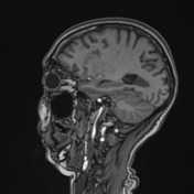
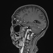
The superior ophthalmic veins are bilaterally enlarged although there is no evidence of cavernous sinus thrombosis. The remainder of the dural venous sinuses are patent. There is dilatation of the optic nerve sheaths bilaterally.
Likely small left cerebellar hemisphere developmental venous anomaly.
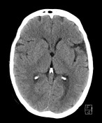
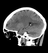
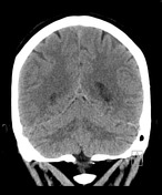

Selected images of CT showing the appearances of the likely pus layering within the posterior horns of the lateral ventricles.
Case Discussion
This case illustrates layering material within the lateral ventricles that almost certainly represents pus, even though there is no appreciable leptomeningeal or ependymal enhancement, which would be expected with this amount of pus.
Dilatation of the optic nerve sheaths bilaterally which can be seen in a setting of increased intracranial pressures, as can dilatation of the superior ophthalmic veins.
Blood Cultures:
Positive culture bottle(s) 1. Streptococcus pneumoniae
Pneumococcal meningitis/ventriculitis has been suspected based on the blood culture.


 Unable to process the form. Check for errors and try again.