Presentation
Child attends for presumptive posterior triangle lump. Incidental finding on MRI.
Patient Data


Well defined T2 hyperintensive cystic lesion in the floor of the mouth.
No solid components.
Separate from the tongue.

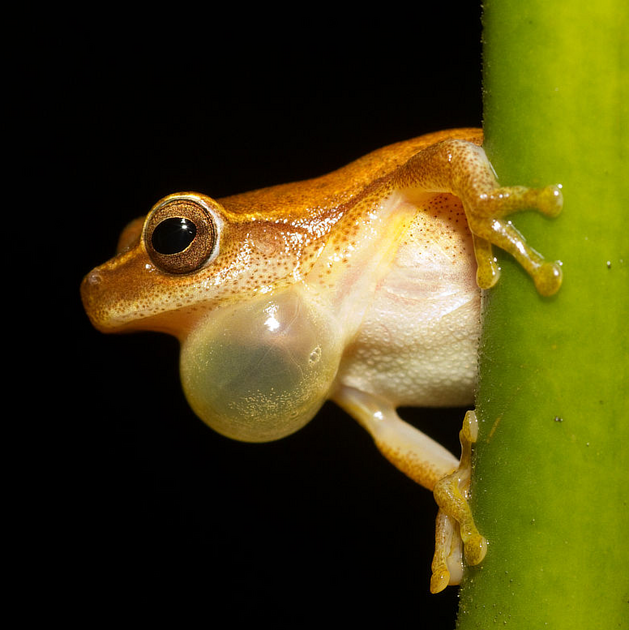
Dendropsophus microcephalus. Photo by Brian Gratwicke. See case discussion for full attribution.
Case Discussion
The term ranula is derived from the Latin word for frog. It describes the cystic space beneath the tongue being like the pouch/underbelly of a frog. It represents a mucocele in the floor of the mouth. The MRI signal within it is typically high on T2-weighted images due to the mucin content.
Attribution
Original file: https://en.wikipedia.org/wiki/File:Dendropsophus_microcephalus_-_calling_male_(Cope,_1886).jpg
Author: User:Brian.gratwicke (Brian Gratwicke at Flickr)
License: CC BY 2.0


 Unable to process the form. Check for errors and try again.