
Presentation
Frontal headache and blurring of vision
Patient Data
Age: 35 years
Gender: Male
From the case:
Rathke cleft cyst

Show annotations
Download
Info

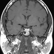
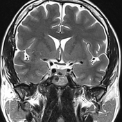
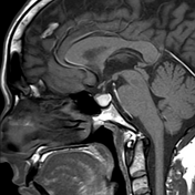
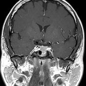
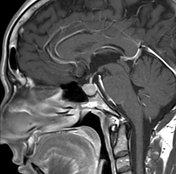
A well-defined sellar cystic lesion exhibits high signal intensity on T1 and intermediate signal in T2 with an intracystic hypointense nodule. It shows no enhancement in the post-contrast study apart from minimal marginal enhancement.
Case Discussion
Here is a case of Rathke cleft cyst with an intracystic nodule of low signal in T2 WI that is virtually pathognomonic. This patient has been under MRI follow up for 5 years with no detectable changes of this lesion


 Unable to process the form. Check for errors and try again.