
Presentation
Headache
Patient Data


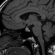
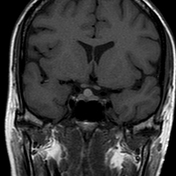
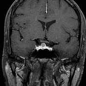
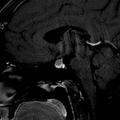
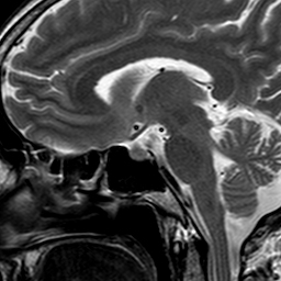
Well defined rounded lesion centered within the midline of anterior pituitary gland involving pituitary stalk showing T2 hypointense signal, T1 hyperintense signal without post contrast enhancement. Lesion measures 8 x 8 x 7 mm. There is no evidence of invasion of the cavernous sinuses. Optic chiasma is normal and is undisplaced.
The anterior pituitary normal in height. Posterior pituitary bright spot is seen and is in normal location.
Findings are likely suggestive of Rathkes cleft cyst
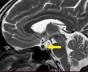
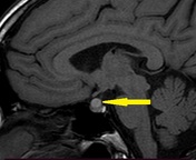
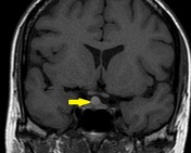
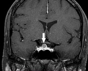
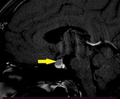
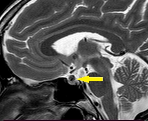
Annotated images showing pituitary lesion.
Case Discussion
Patient came with complaints of headache. Pituitary hormonal studies were normal. No treatment was taken.
Imaging differential include- Pituitary microadenoma
Follow up imaging after 1 year showed stable findings.


 Unable to process the form. Check for errors and try again.