
Presentation
Hyperprolactinemia.
Patient Data


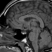
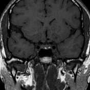

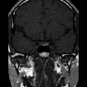
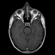
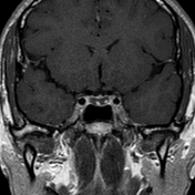
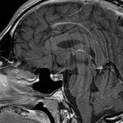
A well-defined sellar cyst elicits a low signal onT1 and a high signal on T2 with intracystic two small nodules eliciting a high signal on T1 and a low signal on T2 WI. The cyst shows rim enhancement on the post-contrast series. Central position of the pituitary infundibulum. There is no extension into the cavernous sinuses and the cavernous carotid flow voids are maintained.
Conclusion: The imaging features with typical intracystic nodules are pathognomonic of Rathke's cleft cyst.
Case Discussion
The patient had a stationary size and signal of the cyst and the intracystic nodules on repeated MRI studies for two years.
In Rathke cleft cysts, about75% of cases might show a small non-enhancing intracystic nodule which is virtually pathognomonic of a Rathke cleft cyst. It is usually hyperintense to surrounding fluid on T1 and hypointense on T2. Depending on the signal of the surrounding fluid, it may be inapparent on one sequence or the other.


 Unable to process the form. Check for errors and try again.