
Presentation
Rectal bleeding.
Patient Data
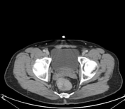


Segmental circumferential wall thickening of the rectum with associated few small mesorectal lymph nodes.
No hepatic deposits or peritoneal lesions are identified.
A colonoscopy and biopsy were performed.
Histopathology
Microscopic:
Sections show a moderately differentiated gland-forming carcinoma with desmoplasia. The glands are filled with necrotic debris (dirty necrosis), and inflammatory cells are scattered. Tubules range from simple and complex to slightly irregular. Mitoses are frequent. In the background, there is evidence of an underlying low-grade villous adenoma.
Diagnosis:
Rectal mass; colonoscopic biopsy.
Invasive adenocarcinoma, grade II, arising in a low-grade villous adenoma.
Case Discussion
Early detection of malignant lesions is a critical responsibility in radiology. A meticulous and thorough evaluation of bowel segments is essential in all abdominal imaging studies, particularly in cases with a high-risk clinical history, such as this one.
This approach ensures the timely identification of potential pathology and guides appropriate clinical management.


 Unable to process the form. Check for errors and try again.