
Presentation
Palpable non-painful right abdominal wall mass noticed 10 days ago.
Patient Data
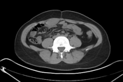


Focal enlargement of the right rectus abdominis muscle caused by a well-defined homogenous intramuscular lesion that appears relatively isodense to the surrounding muscles with no calcifications, fat, or areas of cystic degeneration inside. No evidence of stranding of the adjacent fat or invasion of the adjacent structures.

A well-circumscribed oval hypoechoic mass seen within the right rectus abdominis muscle showing no internal vascularity on Doppler scan & measures about 1.9 x 3.8 x 5.8 cm in Ap x T x ht.
Case Discussion
This young woman noticed this non-painful mass in the right paraumbilical region about 10 days before the examination.
Radiological findings are highly suggestive of rectus abdominis desmoid tumour.
Unfortunately, the pathology of this lesion isn't available and other differential diagnoses should be included like lymphoma, sarcoma, schwannoma and metastasis.
Desmoid tumours are benign fibroelastic tumours with potential local invasion and recurrence after excision.
A large percentage of desmoid tumours were found to arise more in females in and around pregnancy. They most commonly occur in the anterior abdominal wall muscles, particularly the right rectus abdominis muscle & this may be related to trauma from abdominal stretching and fetal movement 1.


 Unable to process the form. Check for errors and try again.