
Presentation
Incidental renal cystic mass on ultrasound exam (not shown).
Patient Data
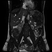

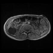
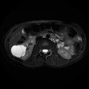
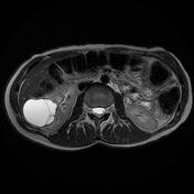
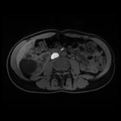
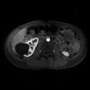
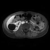
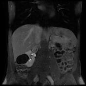

Well-marginated exophytic cystic mass (10 cm) arising from the lower third of the right kidney. It displays a homogeneous fluid content of a low signal on T1, high signal on T2, T2 fat sat and FIESTA with a few thin internal septa (thickness <2 mm). No evidence of peripheral or septal enhancement on postcontrast sequences.
Case Discussion
MRI features most consistent with a renal cyst class II according to the Bosniak classification system of renal cystic masses.
Renal cystic masses are commonly encountered in our daily practice. The Bosniak classification system for renal cystic masses is helpful in determining the class of the cyst and therefore its management.
A Bosniak classification, version 2019 has been proposed to increase the accuracy and include MRI features but does not yet (2022) have widespread validation.


 Unable to process the form. Check for errors and try again.