Presentation
Ventricular tachycardia for ICD insertion.
Patient Data


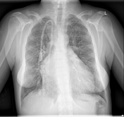
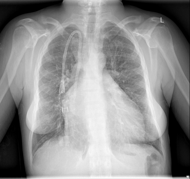
Cardiomegaly.
Dual-lumen venous catheter tip at the cavo-atrial junction.
Clear lungs and pleural spaces.
Erosion at the distal end of the clavicles with widened A-C joints.
Surgical clips in the base of the neck.
Otherwise normal.


Small kidneys containing multiple renal cysts.
Osteodystrophy with abnormal bone texture and marked osteosclerosis sparing the mid vertebral bodies, rugger-jersey spine appearance.
Case Discussion
The diagnosis of hyperparathyroid bone disease was suspected from the characteristic clavicular erosion and surgical clips. A full history was obtained from the medical records:
end-stage kidney disease due to multicystic dysplastic kidney, somatic mutation
on peritoneal dialysis until membrane failure, then converted to haemodialysis, total time five years
total parathyroidectomy for secondary hyperparathyroidism
idiopathic cardiomyopathy with normal coronary arteries diagnosed while in ICU for severe pneumonia and decompensated heart failure
work-up for simultaneous heart/kidney transplant


 Unable to process the form. Check for errors and try again.