
Presentation
Right loin pain in a diabetic patient. The ultrasound exam (not shown) revealed an enlarged hypoechoic right kidney.
Patient Data
Age: 40 years
Gender: Female
From the case:
Renal vein thrombosis


Show annotations
Download
Info
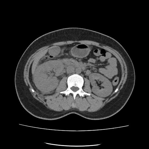
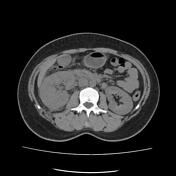
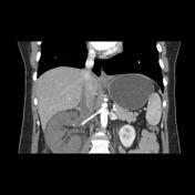
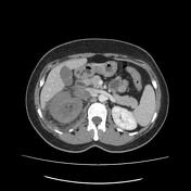
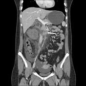
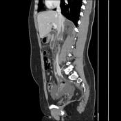
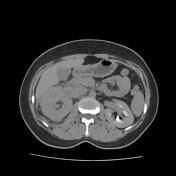
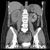
The CT scan demonstrates enlarged right kidney with:
lack of cortical and parenchymal enhancement on arterial and venous phase
absent calyceal opacification on excretory phase
stranding of the perinephric fat
thickening of the peritoneal reflections
enlarged right renal vein with a filling defect during the venous phase, extending to the IVC
the left renal vein and renal arteries are patent
mild to moderate intraperitoneal effusion
Case Discussion
CT features of a right renal vein thrombosis extending to the IVC with enlarged hypodense non-enhancing kidney in a diabetic patient.


 Unable to process the form. Check for errors and try again.