
Presentation
Three months history of visual floaters with progressive reduced visual acuity
Patient Data

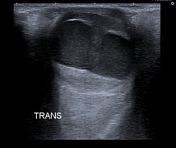
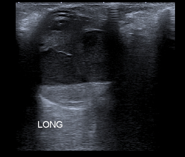
The ultrasound images show a retinal detachment with a fluid-fluid level (hemorrhage).

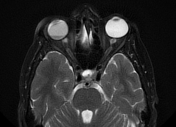
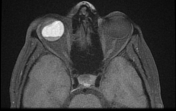
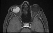
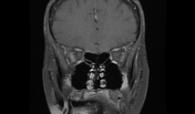

The MRI sequences demonstrate an extensive retinal detachment with a typical V-shape with an anterior insertion into the ora Serrata (well-visualized on T2 fat saturation) and posterior insertion into the optic disc. A subretinal hemorrhage is noted of high signal on T1WI/FLAIR, low signal on T2WI with fluid-fluid level (the hypointense sediment on all sequences indicates blood products). No choroidal lesion is seen.
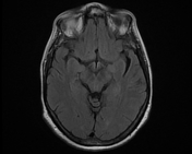
Normal MR appearance of the left ocular globe and optic nerves.
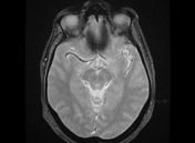
Asymmetrical size of the lacrimal glands with no focal lesion seen.
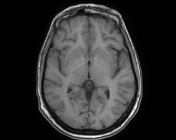
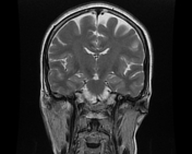
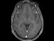
Normal appearance of the posterior cerebral fossa and supratentorial region.
Case Discussion
Ultrasound and MRI features are most consistent with a retinal detachment.
On imaging, the differential diagnosis includes:


 Unable to process the form. Check for errors and try again.