
Presentation
Painless progressive loss of vision in the left eye. No history of trauma.
Patient Data
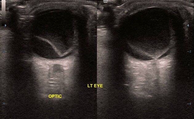
A thin, smooth, and V-shaped freely floating membrane is noted extending from the optic disc to the ora serrata, typical of retinal detachment. There is mildly echogenic debris within the posterior chamber, consistent with recent vitreous haemorrhage. No tumours or retinal tears are seen. The lens is intact with no internal echoes.
The right eye shows normal imaging features.
Case Discussion
These features are typical of a recent total retinal detachment.
Ultrasonography plays a vital role in the diagnosis of retinal detachment, especially in cases with opaque media. Ultrasound evaluation can provide valuable information about the subretinal space to rule out tumours, which may be obscured on ophthalmoscopic examination.
Retinal detachment is caused by trauma, tumours, and other disease processes such as diabetic retinopathy. Determination of the cause and severity of the detachment plays a major role in patient management. Acute detachments are manageable, but if left untreated, they may become inoperable and irreversible.


 Unable to process the form. Check for errors and try again.