
Presentation
Headache.
Patient Data
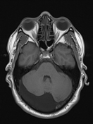


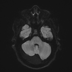
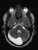


A large right retro-cerebellar extra-axial cyst is seen compressing the right cerebellar hemisphere and elevating the tentorium being insinuated between the occipital lobes. It is seen following CSF signal on all sequences with facilitated diffusion. It is seen measuring 3.2X7.7X6.5 cm along its axial and cranio-caudal dimensions.
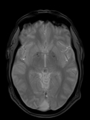
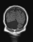
Annotated image showing the membrane of the arachnoid cyst (red arrows). Remodeling of the skull is also present (blue arrow head).


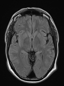
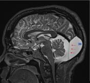
Postoperative status after shunt tube placement within the aforementioned cystic lesion seen running along the posterior surface of right cerebellar hemisphere (cystoperitoneal shunt tube).
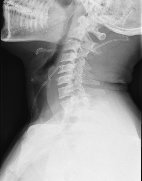
X- ray images following the course of the cystoperitoneal shunt tube seen extending from posterior cranial fossa down to the pelvic cavity. No evidence of shunt break or dislodgement could be detected.
Case Discussion
This case illustrates typical features of an arachnoid cyst, with visualization of the cyst membrane and bone remodeling. Also, it shows cysto-peritoneal shunting which is a therapeutic option for treatment of symptomatic arachnoid cyst.


 Unable to process the form. Check for errors and try again.