Presentation
Abdominal pain for investigation. Incidental anatomical variant identified.
Patient Data
Age: 85 years
Gender: Female
From the case:
Retroaortic left renal vein
Download
Info
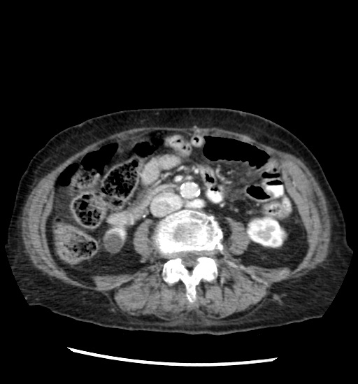
- retroaortic left renal vein
- double right renal veins emptying directly into the inferior vena cava
- cyst at the inferior pole of the right kidney
Case Discussion
Retroaortic left renal vein is an anatomic variant typically identified as an incidental finding on imaging. Whilst usually asymptomatic, a retroaortic left renal vein can be compressed by the aorta and cause venous congestion of the kidney or left gonadal vein, leading to varicocele (male) or pelvic congestion syndrome (female). A retroaortic left renal vein is also important to note prior to performing any renal/retroperitoneal surgery.