
Presentation
The primary complaint was a reducible umbilical protrusion for seven months. Ultrasound incidentally revealed a huge mass, CT was done later on.
Patient Data
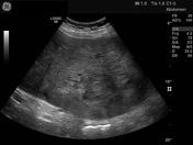
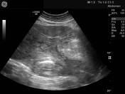

Inhomogenous intra-abdominal huge mass with degenerative calcifications.
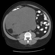

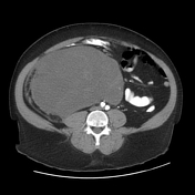
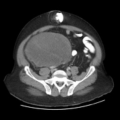
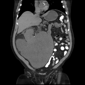
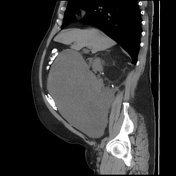

There is right side subhepatic huge retroperitoneal fat/solid mass with septations and dystrophic calcifications and thickened septations and a large solid component that has some areas of cystic degeneration. The mass displaces the intra-abdominal structures including the right kidney, liver and gut. There is also mass induced umbilical hernia and a right-sided inguinal omentocele.
Case Discussion
Sagittal and axial images demonstrated clear separation between the mass and the kidney excluding angiomyolipoma (AML). Also, excess lipid components excluded neurogenic tumors like schwannoma. In addition, the presence of calcification supported the diagnosis of liposarcoma rather than lipomyosarcoma, in which calcifications are a rare finding.


 Unable to process the form. Check for errors and try again.