
Presentation
Worsening pain in hands: osteoarthritis versus rheumatoid arthritis
Patient Data

No previous imaging available for comparison.
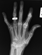
Right hand: ankylosis of the carpus and 2nd-3rd carpometacarpal joints. Mild loss of joint space at the remaining carpometacarpal joints, radiocarpal joints, and distal radioulnar joint. Positive ulnar variance noted. Resorption of the ulnar styloid process. Multiple subchondral cysts seen within the distal radius. Partial ankylosis with subluxations and erosions at the 3rd-5th metacarpophalangeal joints. Ankylosis at the proximal interphalangeal joint of the index finger, with probable boutonniere deformity. Imaging features are in keeping with advanced rheumatoid arthritis.
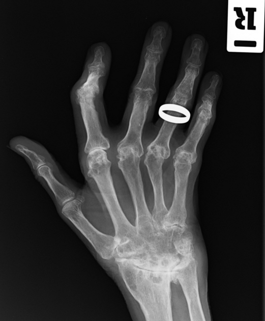
Left hand: ankylosis of the carpus and 2nd-5th carpometacarpal joints. Marked loss of joint space seen at the radiocarpal joint, distal radioulnar joint, and first carpometacarpal joint. Complete resorption of the ulnar styloid process. Subchondral cyst formation within the distal radius. Marked loss of joint space with erosions at the head of the second and third metacarpophalangeal joints. Ankylosis at the proximal interphalangeal joint of the index finger, with probable boutonniere deformity. Imaging features are in keeping with advanced rheumatoid arthritis.
Case Discussion
Advanced rheumatoid arthritis.


 Unable to process the form. Check for errors and try again.