
Presentation
Follow-up imaging after surgical resection of the nasal cavity and paranasal sinuses fungal infection in a patient with a long history of poorly controlled diabetes mellitus and recent headaches.
Patient Data


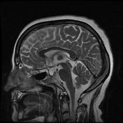
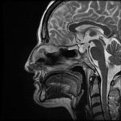

Post-treatment changes at nasal cavity or paranasal sinuses
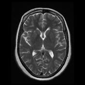
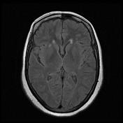
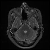
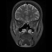
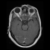
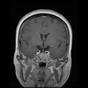
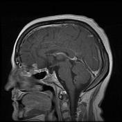
Two abnormal signal ring-enhancing lesions at orbitofrontal cortex just above the cribriform plate measuring about 10 mm and 7 mm on the left and right sides respectively due to intracranial extension of the recent known sinonasal fungal infection
Mild mucosal thickening also is seen at paranasal sinuses
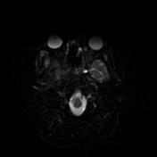
Intraconal low signal intensity (on T1/T2) heterogenous enhancing mass-like lesion near the left orbital apex with adhesion to optic nerve sheath could be due to infiltrative or infective processes.
High signal foci in T2 and flair sequences at subcortical and periventricular white matter of both cerebral hemispheres depict microvascular ischaemic events.
Case Discussion
With regarding the imaging findings, intracranial extension of the known sinonasal fungal infection (rhinocerebral mucormycosis) is the final diagnosis.


 Unable to process the form. Check for errors and try again.