
Presentation
Maternal history of vitamin D deficiency, child unable to properly stand, clinical query of metabolic bone disease.
Patient Data

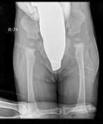

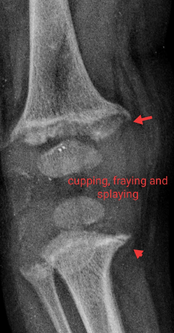
There is cupping, fraying and irregularity at distal femoral, proximal and distal tibial and fibular metaphyses with widening of physes bilaterally. Similar changes of lesser magnitude are also present at the proximal femoral metaphysis on both sides. Overall these described imaging appearances are suggestive of rickets. Serum vitamin D levels are recommended. There is no evidence of clinically-suspected developmental dysplasia of the hip or Blount disease. No fracture or dislocation is present. Normal soft tissue shadows.
Additional contributor: Assistant Professor Dr Jaideep Dareera
Case Discussion
Radiographic features of rickets lag behind biochemical and clinical improvements by about two weeks. Harris growth arrest lines are dense lines traversing parallel to the metaphyses which may be used as a marker of old rickets.


 Unable to process the form. Check for errors and try again.