
Presentation
10 day history of flu symptoms and productive cough.
Patient Data

Findings:
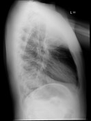
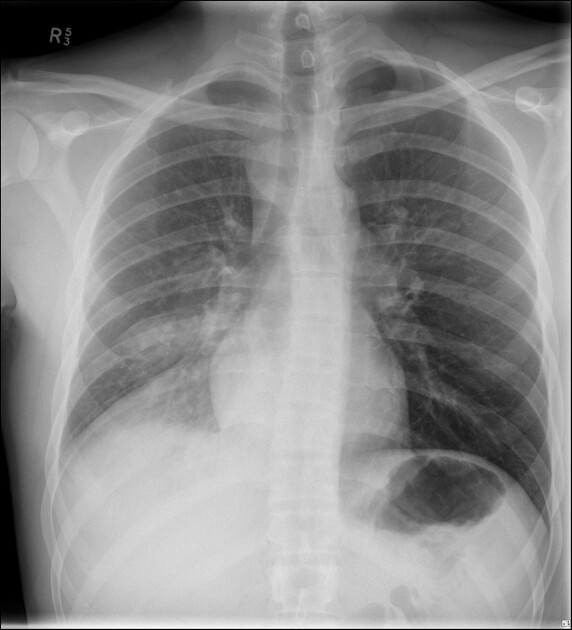
The right hemidiaphragm is obscured and there is overlying consolidation with air bronchograms reflecting right lower lobe consolidation. There is a probable small right sided pleural effusion. The left lung is well expanded and is clear. The cardiomediastinal silhouette is within normal limits.
Conclusion:
Right lower lobe pneumonic consolidation with a probable small right pleural effusion.
Case Discussion
This case demonstrates one of the many manifestations of the silhouette sign on chest radiographs. There is right lower lobe consolidation with loss of the contour of the right hemidiaphragm. An infective etiology is the likely cause given the history and the chest x-ray findings, with pneumonia being the primary differential diagnosis.


 Unable to process the form. Check for errors and try again.