
Presentation
Chronic cough and exertional dyspnea. Chest x-ray performed.
Patient Data
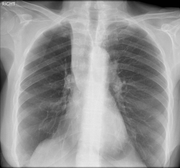
Linear density medially in the right upper lobe.
Volume loss in the right hemithorax.
Relative translucency of the right hemithorax.
Elevated right hilum.
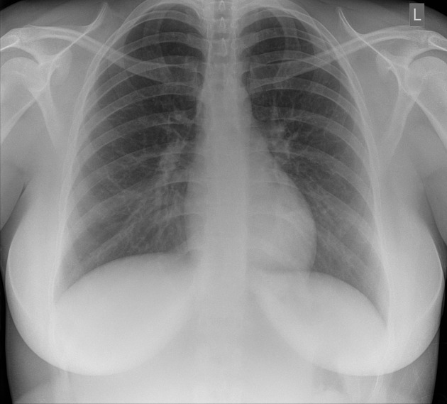
A normal female chest x-ray for comparison with the first film.
Case Discussion
Benjamin Felson was the doyan of chest radiology and his description of the silhouette sign remains a core radiological plain film lesson.
The collapse or consolidation of the various lobes of the lung illustrates classical appearances - one of these being the right upper lobe.
The right upper lobe will collapse medially effacing the upper mediastinum.
This film has a number of accompanying features:
a. The transradiancy of the right lung ( compared to the left ) is greater - it reflects the hemithorax now being only occupied by the middle and lower lobes.
b. Reduction in the hemithorax volume
c. Elevation of the right hilum ( it is usually 1.5cm lower than the left, but in this case at the same level as it is 'pulled up' )
The features are even better appreciated if compared with the normal chest x-ray which is shown above.


 Unable to process the form. Check for errors and try again.